Catalog # |
Size |
Price |
|
|---|---|---|---|
| FR-056-52 | 1 nmol | $382 |
|
| Exhibits correct molecular weight |
|
| Each vial contains 1 nmol of NET labeled peptide. |


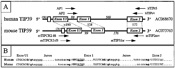
A, Schematic representation of the known portions of the human (upper panel) and the mouse (lower panel) TIP39 gene. The names of the different exons are indicated; the sizes of exons (normal letters) and introns (italic letters) are given in base pairs; the approximate positions of the different PCR primers are shown (see Materials and Methods); note that the positions of the universal AP1 and AP2 primers that were used for 5' RACE are arbitrary. B, Splice donor/acceptor sites in the human and mouse gene are shown; exonic nucleotides are shown in capital letters; intronic nucleotides in lowercase letters; splice site consensus nucleotides are in bold; the initiator ATG in exon 1 is underlined.
Tuberoinfundibular peptide of 39 residues (TIP39) was identified as the endogenous ligand of parathyroid hormone 2 receptor. We have recently demonstrated that TIP39 expression in adult rat brain is confined to the subparafascicular area of the thalamus with a few cells extending laterally into the posterior intralaminar thalamic nucleus (PIL), and the medial paralemniscal nucleus (MPL) in the lateral pontomesencephalic tegmentum. During postnatal development, TIP39 expression increases until postnatal day 33 (PND-33), then decreases, and almost completely disappears by PND-125. Here, we report the expression of TIP39 during early brain development. TIP39-immunoreactive (TIP39-ir) neurons in the subparafascicular area first appeared at PND-1. In contrast, TIP39-ir neurons were detectable in the MPL at embryonic day 14.5 (ED-14.5), and the intensity of their labeling increased thereafter. We also identified TIP39-ir neurons between ED-16.5 and PND-5 in two additional brain areas, the PIL and the amygdalo-hippocampal transitional zone (AHi). We confirmed the specificity of TIP39 immunolabeling by demonstrating TIP39 mRNA using in situ hybridization histochemistry. In the PIL, TIP39 neurons are located medial to the CGRP group as demonstrated by double immunolabeling. All TIP39-ir neurons in the AHi and most TIP39-ir neurons in the PIL disappear during early postnatal development. The adult pattern of TIP39-ir fibers emerge during postnatal development. However, fibers emanating from PIL can be followed in the supraoptic decussations towards the hypothalamus at ED-18.5. These TIP39-ir fibers disappear by PND-1. The complex pattern of TIP39 expression during early brain development suggests the involvement of TIP39 in transient functions during ontogeny.
Brenner D, Bagó AG, Gallatz K, Palkovits M, Usdin TB, Dobolyi A. Tuberoinfundibular peptide of 39 residues in the embryonic and early postnatal rat brain. J Chem Neuroanat. 2008;36(1):59-68.
By screening public databases, we identified human and mouse genomic DNA clones that encode the tuberoinfundibular peptide of 39 residues (TIP39). The TIP39 precursor is encoded by at least three exons; a noncoding exon U1, exon 1 encoding residues ?61 (initiator methionine) to ?19 of the leader sequence, and exon 2 encoding residues ?18 to ?1 and residues +1 to +39. Secreted human TIP39 is identical to the previously isolated bovine TIP39, whereas mouse TIP39 differs by four amino acids. Phylogenetic analyses suggested that TIP39, PTH, and PTHrP may have evolved from a common ancestor. Synthetic human and mouse TIP39 showed indistinguishable potencies [EC50: 0.54 (human) vs. 0.74 nm (mouse)] at the human PTH2-receptor stably expressed in LLCPK1 cells; furthermore, TIP-(9–39) was an inhibitor of cAMP accumulation stimulated by either [Tyr34]PTH(1–34)amide or human/bovine TIP39. In the mouse, an approximately 4.5-kb mRNA encoding TIP39 was identified by Northern blot analysis in testis and, less abundantly, in liver and kidney, whereas other tissues revealed additional smaller transcripts. In situ hybridizations revealed TIP39 expression in seminiferous tubuli and several brain regions, including nucleus ruber, nucleus centralis pontis, and nucleus subparafascicularis thalami. Because PTH2 receptor expression was previously shown to be highest in brain, pancreas, and testis, our findings are consistent with the notion that TIP39 is a neuropeptide which may also have a role in spermatogenesis.
John MR, Arai M, Rubin DA, Jonsson KB, Jüppner H. Identification and characterization of the murine and human gene encoding the tuberoinfundibular peptide of 39 residues. Endocrinology. 2002;143(3):1047-57.
Tuberoinfundibular peptide is a recently discovered agonist for the PTH receptor-2; the latter has a wide distribution including the external zone of the median eminence of the hypothalamus, suggesting a role in neuroendocrine function. We have investigated the effects of tuberoinfundibular peptide on the hypothalamo-pituitary axes in vitro and in vivo. Tuberoinfundibular peptide had effects on the hypothalamo-pituitary-adrenal axis with increased release of ACTH-releasing factor (tuberoinfundibular peptide 100 nM 4.4 +/- 0.6 pmol/explant vs. control 2.9 +/- 0.4 pmol/explant, P < 0.001) and increased release of arginine vasopressin (tuberoinfundibular peptide 100 nM 563.5 +/- 55.5 fmol/explant vs. control 73.4 +/- 9.6 fmol/explant, P < 0.01) from in vitro hypothalamic explants. Intracerebroventricular administration of tuberoinfundibular peptide and PTH((1-34)) resulted in elevated plasma ACTH at 10 min post injection (saline 13.5 +/- 2.1 pg/ml, tuberoinfundibular peptide 3 nmol 32.3 +/- 4.0 pg/ml; P < 0.01 to saline: PTH((1-34)) 10 nmol 28.9 +/- 3.2 pg/ml: P < 0.05 to saline). Tuberoinfundibular peptide also had both in vitro and in vivo effects on the hypothalamo-pituitary-gonadal axis with increased release of LH-releasing hormone (tuberoinfundibular peptide 100 nM 28.5 +/- 5.1 fmol/explant vs. control 19.3 +/- 2.5 fmol/explant, P < 0.05) from in vitro hypothalamic explants. Both intracerebroventricular and peripheral administration of tuberoinfundibular peptide had effects on the hypothalamo-pituitary-gonadal axis. Intracerebroventricular injection of tuberoinfundibular peptide increased plasma LH (tuberoinfundibular peptide 10 nmol 0.70 +/- 0.09 ng/ml vs. saline 0.42 +/- 0.04 ng/ml at 10 min, P < 0.05). Intraperitoneal administration of tuberoinfundibular peptide also increased plasma LH (tuberoinfundibular peptide 30 nmol 0.53 +/- 0.09 ng/ml vs. saline 0.21 +/- 0.04 ng/ml at 10 min, P < 0.05). In addition to these actions on the hypothalamo-pituitary-adrenal and hypothalamo-pituitary-gonadal axes, an increased release of GH-releasing factor (GRF) from hypothalamic explants (tuberoinfundibular peptide 100 nM 770.9 +/- 90.7 pg/explant vs. control 657.8 +/- 77.7 pg/explant, P < 0.01) was observed. Overall, these data show the actions of tuberoinfundibular peptide on the hypothalamo-pituitary axes and suggest that it may play a role in the control of the hypothalamo-pituitary-adrenal and hypothalamo-pituitary-gonadal axes.
Ward HL, Small CJ, Murphy KG, Kennedy AR, Ghatei MA, Bloom SR. The actions of tuberoinfundibular peptide on the hypothalamo-pituitary axes. Endocrinology. 2001;142(8):3451-6.
|
|
Phylogenetic analysis indicating the evolutionary relationship among precursor proteins of the TIP39, PTH, PTHrP, and secretin families of peptides. A Neighbor-Joining phylogenetic analysis using distance as the criteria is shown above (tree length = 740, consistency index excluding uninformative residues = 0.876, with 165 parsimony-informative characters). The bootstrap/jackknife values from 10,000 replicates indicate support of a given node where 95% is considered to be significant (36 37 ). A Maximum Parsimony analysis using parsimony as the criteria generated a similar phylogenetic relationship between PTH, PTHrP, TIP39, secretin, and GIP (tree length = 746, consistency index excluding uninformative residues = 0.846, with 165 parsimony-informative characters). In addition to the Neighbor-Joining and Maximum Parsimony phylogenetic analyses, Quartet puzzling using Maximum Parsimony criteria and Star-decomposition (tree length = 739, consistency index excluding uninformative characters = 0.855, with 165 parsimony-informative characters) (35 36 ) support the hypothesis that PTH and PTHrP are sister groups, and that TIP39 is the sister group to this clade. PTH, PTHrP, and TIP39 thus form a superfamily, whereas secretin and VIP (not shown) appears to be a sister group to this larger superfamily (accession nos.: PTH (cat, AF309967; chick, M36522; cow, J00024; dog, U15662; horse, AF134233; human, NM_000315; macaque, AF130257; mouse, NM_020623; pig, X05722; and rat, NM_017044); PTHrP (chick, X52131; dog, U15593; cow, P58073; human, J03580; mouse, M60056; rabbit, AF219973; rat, NM_012636; sheep, AF327654; fugu, AJ249391; sparus, AF197904); VIP (chick, U09350; mouse AK018599; human XM_004381); secretin [mouse, X73580; pig, M31496; human, XM_012014; and human GIP (NM_004123)]. Markus R. et al. Endocrinology Vol. 143, No. 3 1047-1057 (2002). |
Hypothalamic releasing factors released from medial basal hypothalamic explants |
||||||||||||||||||||||||||||||||||
|
||||||||||||||||||||||||||||||||||
|
Basal, TIP39 100 nM followed by potassium 56 mM incubation periods each of 45 min duration. Values are shown as mean ± SEM. 1 P < 0.001, 2 P < 0.01, 3 P < 0.05, by t test.
H. L. Ward, et al. Endocrinology Vol. 142, No. 8 3451-3456 (2001)
|
||||||||||||||||||||||||||||||||||
No References
| Catalog# | Product | Size | Price | Buy Now |
|---|
Social Network Confirmation