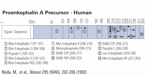
Skin responds to environmental stressors via coordinated actions of the local neuroimmunoendocrine system. Although some of these responses involve opioid receptors, little is known about cutaneous proenkephalin expression, its environmental regulation, and alterations in pathology. The objective of this study was to assess regulated expression of proenkephalin in normal and pathological skin and in isolated melanocytes, keratinocytes, fibroblasts, and melanoma cells. The proenkephalin gene and protein were expressed in skin and cultured cells, with significant expression in fibroblasts and keratinocytes. Mass spectroscopy confirmed Leu- and Met-enkephalin in skin. UVR, Toll-like receptor (TLR)4, and TLR2 agonists stimulated proenkephalin gene expression in melanocytes and keratinocytes in a time- and dose-dependent manner. In situ Met/Leu-enkephalin peptides were expressed in differentiating keratinocytes of the epidermis in the outer root sheath of the hair follicle, in myoepithelial cells of the eccrine gland, and in the basement membrane/basal lamina separating epithelial and mesenchymal components. Met/Leu-enkephalin expression was altered in pathological skin, increasing in psoriasis and decreasing in melanocytic tumors. Not only does human skin express proenkephalin, but this expression is upregulated by stressful stimuli and can be altered by pathological conditions.
This publication used Leu-enkephalin EIA kit (EK-024-21) from Phoenix Pharmaceuticals to measure the blood level for Leu-Enkephalin.
Slominski AT, Zmijewski MA, Zbytek B, et al. J Invest Dermatol. 2011;131(3):613-22.
OBJECTIVE: To assess mean daily plasma concentrations of ACTH, cortisol, DHEAS, leu-enkephalin, and beta-endorphin in epilepticpatients with complex partial seizures evolving to tonic-clonic in relation to frequency of seizure occurrence (groups with seizure occurrences - several per week and several per year) and duration of the disease (groups less than and more than 10 years). We decided to analyse mean daily values of beta-endorphin and leu-enkephalin because of significant differences in concentrations of these substances in blood during the day.MATERIAL AND METHODS: The study was performed on 17 patients (14 males + 3 females; mean age 31.8 yrs) treated with carbamazepine (300-1800 mg/day). The control group consisted of six age-matched healthy volunteers. Blood was collected at 8 a.m., 2 p.m., 8 p.m., and 2 a.m. Intergroup analysis was performed with the use of ANOVA Kruskal-Wallis test.RESULTS: Mean daily concentrations of ACTH and cortisol in the blood of the patients with epilepsy were higher in comparison with those of the healthy volunteers, independently of the frequency of seizures and duration of the disease. Mean daily concentrations of beta-endorphinin the blood of the patients with epilepsy were higher in the groups of patients with more severe clinical course of disease (with more frequently occurring epilepsy seizures and longer duration of the disease) in comparison with healthy subjects. Mean daily concentrations of leu-enkephalin in the blood of the patients with epilepsy were higher in the group of patients with short duration of the disease in comparison with the group with long duration of the disease.CONCLUSIONS: 1. Pituitary-adrenal axis hyperactivity is observed in patients with clinically active epilepsy, independently of the frequency of seizures and duration of the disease. 2. Changes in endogenous opioid system activity are related to the clinical activity of epilepsy - beta-endorphin concentrations are connected with frequency of seizures and duration of the disease and leu-enkephalin concentrations with duration of the disease. 3. Endogenous opioid peptides might take part in the neurochemical mechanism of human epilepsy.This publication used Leu-enkephalin RIA kit (RK-024-21) from Phoenix Pharmaceuticals to measure the blood level for Leu-Enkephalin.
Marek B, Kajdaniuk D, Kos-kud?a B, et al. Endokrynol Pol. 2010;61(1):103-10.
Neprilysin (NEP) is a zinc metallopeptidase that efficiently degrades the amyloid beta (Abeta) peptides believed to be involved in the etiology of Alzheimer disease (AD). The focus of this study was to develop a new and tractable therapeutic approach for treating AD using NEP gene therapy. We have introduced adeno-associated virus (AAV) expressing the mouse NEP gene into the hindlimb muscle of 6-month-old human amyloid precursor protein (hAPP) (3X-Tg-AD) mice, an age which correlates with early stage AD. Overexpression of NEP in muscledecreased brain soluble Abeta peptide levels by approximately 60% and decreased amyloid deposits by approximately 50%, with no apparent adverse effects. Expression of NEP on muscle did not affect the levels of a number of other physiological peptides known to be in vitro substrates. These findings demonstrate that peripheral expression of NEP and likely other peptidases represents an alternative to direct administration into brain and illustrates the potential for using NEP expression in muscle for the prevention and treatment of AD.
This publication used Leu-enkephalin EIA kit (EK-024-21) from Phoenix Pharmaceuticals to measure Leu-Enkephalin level in different regions of transgenic mouse brain.
Liu Y, Studzinski C, Beckett T, et al. Mol Ther. 2009;17(8):1381-6.
We have reported that transplantation of adrenal medullary chromaffin cells that release endogenous opioid peptides into pain modulatory regions in the CNS produce significant antinociceptive effects in patients with terminal cancer pain. However, the usefulness of this procedure is minimal because the availability of human adrenal tissue is very limited. Alternative xenogeneic materials, such as porcine and bovine adrenal chromaffin cells present problems of immune rejection and possible pathogenic contamination. In an attempt to develop opioid peptide-producing cells of autologous origin, we have transfected human mesenchymal stem cells (hMeSCs) with a mammalian expression vector containing a fusion gene of green fluorescent protein (GFP) and human preproenkephalin (hPPE), a precursor protein for enkephalin opioid peptides. Enkephalins are major neurotransmitters that play an important role in analgesia by activating peripheral opioid receptors. Following the establishment of stable transfection of hMeSCs, the expressions of hPPE and GFP were confirmed and the production of methionine enkephalin (Met-enkephalin) was significantly increased compared to control naive hMeSCs (p < 0.05). Our in vitro data demonstrated that genetically engineered hMeSCs with transfected hPPE gene can constitutively produce opioid peptide Met-enkephalin at an augmented high level. hMeSCs are relatively easy to isolate from a patient's bone marrow aspirates and expand in culture by repeated passages. Autologous hMeSCs would not require immunosuppression when transplanted back into the same patient. Through targeted gene manipulation such as hPPE gene transfection, this may offer a virtually unlimited safe cell supply for the treatment of opioid-sensitive pain in humans.
This publication used Met-enkephalin peptide (024-35) from Phoenix Pharmaceuticals.
Sugaya I, Qu T, Sugaya K, Pappas GD. Cell Transplant. 2006;15(3):225-30.
The detrimental effect of severe hypoxia (SH) on neurons can be mitigated by hypoxic preconditioning (HPC), but the molecular mechanisms involved remain unclear, and an understanding of these may provide novel solutions for hypoxic/ischemic disorders (e.g. stroke). Here, we show that the delta-opioid receptor (DOR), an oxygen-sensitive membrane protein, mediates the HPC protection through specific signaling pathways. Although SH caused a decrease in DOR expression and neuronal injury, HPC induced an increase in DOR mRNA and protein levels and reversed the reduction in levels of the endogenous DOR peptide, leucine enkephalin, normally seen during SH, thus protecting the neurons from SH insult. The HPC-induced protection could be blocked by DOR antagonists. The DOR-mediated HPC protection depended on an increase in ERK and Bcl 2 activity, which counteracted the SH-induced increase in p38 MAPK activities and cytochrome c release. The cross-talk between ERK and p38 MAPKs displays a "yinyang" antagonism under the control of the DOR-G protein-protein kinase C pathway. Our findings demonstrate a novel mechanism of HPC neuroprotection (i.e. the intracellular up-regulation of DOR-regulated survival signals).
This publication used Leu-enkephalin EIA kit (EK-024-21) from Phoenix Pharmaceuticals to measure Leu-Enkephalin level in culture medium.
Ma MC, Qian H, Ghassemi F, Zhao P, Xia Y. J Biol Chem. 2005;280(16):16208-18.
Intrathecal (i.t.) pretreatment with a low dose (0.3 nmol) of morphine causes an attenuation of i.t. morphine-produced analgesia; the phenomenon has been defined as morphine-induced antianalgesia. The opioid-produced analgesia was measured with the tail-flick (TF) test in male CD-1 mice. Intrathecal pretreatment with low dose (0.3 nmol) of morphine time dependently attenuated i.t. morphine-produced (3.0 nmol) TF inhibition and reached a maximal effect at 45 min. Intrathecal pretreatment with morphine (0.009-0.3 nmol) for 45 min also dose dependently attenuated morphine-produced TF inhibition. The i.t. morphine-induced antianalgesia was dose dependently blocked by the nonselective mu-opioid receptor antagonist (-)-naloxone and by its nonopioid enantiomer (+)-naloxone, but not by endomorphin-2-sensitive mu-opioid receptor antagonist 3-methoxynaltrexone. Blockade of delta-opioid receptors, kappa-opioid receptors, and N-methyl-D-aspartate (NMDA) receptors by i.t. pretreatment with naltrindole, nor-binaltorphimine, and (-)-5-methyl-10,11-dihydro-5H-dibenzo[a,d]cyclohepten-5,10-imine maleate (MK-801), respectively, did not affect the i.t. morphine-induced antianalgesia. Intrathecal pretreatment with antiserum against dynorphin A(1-17), [Leu]-enkephalin, [Met]-enkephalin, beta-endorphin, cholecystokinin, or substance P also did not affect the i.t. morphine-induced antianalgesia. The i.t. morphine pretreatment also attenuated the TF inhibition produced by opioid muagonist [D-Ala2, N-Me-Phe4,Gly-ol5]-enkephalin, delta-agonist deltorphin II, and kappa-agonist U50,488H. It is concluded that low doses (0.009-0.3 nmol) of morphine given i.t. activate an antianalgesic system to attenuate opioid mu-, delta-, and kappa-agonist-produced analgesia. The morphine-induced antianalgesia is not mediated by the stimulation of opioid mu-, delta-, or kappa-receptors or NMDA receptors. Neuropeptides such as dynorphin A(1-17), [Leu]-enkephalin, [Met]-enkephalin, beta-endorphin, cholecystokinin, and substance P are not involved in this low-dose morphine-induced antianalgesia.
This publication used DAMGO peptide (024-10) from Phoenix Pharmaceuticals
Wu HE, Thompson J, Sun HS et al. J Pharmacol Exp Ther. 2004 Jul;310(1):240-6.
This study examined the role of leucine-enkephalin (LE) in the sympathetic regulation of the cardiac pacemaker. LE was administered by microdialysis into the interstitium of the canine sinoatrial node during either sympathetic nerve stimulation or norepinephrine infusion. In study one, the right cardiac sympathetic nerves were isolated as they exit the stellate ganglion and were stimulated to produce graded (low, 20-30 bpm; high 40-50 bpm) increases in heart rate (HR). LE (1.5 nmoles/min) was added to the dialysis inflow and the sympathetic stimulations were repeated after 5 and 20 min of LE infusion. After 5 min, LE reduced the tachycardia during sympathetic stimulation at both low (18.2 +/- 1.3 bpm to 11.4 +/- 1.4 bpm) and high (45 +/- 1.5 bpm to 22.8 +/- 1.5 bpm) frequency stimulations. The inhibition was maintained during 20 min of continuous LE exposure with no evidence of opioid desensitization. The delta-opioid antagonist, naltrindole (1.1 nmoles/min), restored only 30% of the sympathetic tachycardia. Nodal delta-receptors are vagolytic and vagal stimulations were included in the protocol as positive controls. LE reduced vagal bradycardia by 50% and naltrindole completely restored the vagal bradycardia. In Study 2, additional opioid antagonists were used to determine if alternative opioid receptors might be implicated in the sympatholytic response. Increasing doses of the kappa-antagonist, norbinaltorphimine (norBNI), were combined with LE during sympathetic stimulation. NorBNI completely restored the sympathetic tachycardia with an ED50 of 0.01 nmoles/min. A single dose of the micro -antagonist, CTAP (1.0 nmoles/min), failed to alter the sympatholytic effect of LE. Study 3 was conducted to determine if the sympatholytic effect was prejunctional or postjunctional in character. Norepinephrine was added to the dialysis inflow at a rate (30-45 pmoles/min) sufficient to produce intermediate increases (35.2 +/- 1.8 bpm) in HR. LE was then combined with norepinephrine and responses were recorded at 5-min intervals for 20 min. The tachycardia mediated by added norepinephrine was unaltered by LE or LE plus naltrindole. At the same 5-min intervals, LE reduced vagal bradycardia by more than 50%. This vagolytic effect was again completely reversed by naltrindole. Collectively, these observations support the hypothesis that the local nodal sympatholytic effect of LE was mediated by kappa-opioid receptors that reduced the effective interstitial concentration of norepinephrine and not the result of a postjunctional interaction between LE and norepinephrine.
This publication used Leu-enkephalin peptide (024-21) from Phoenix Pharmaceuticals.
Stanfill AA, Jackson K, Farias M, et al. Exp Biol Med (Maywood). 2003;228(8):898-906.
| Catalog# | Product | Standard Size | Price |
|---|---|---|---|
| EK-024-21 | Leu-Enkephalin - EIA Kit | 96 wells | $570 |
| 024-21 | Leu-Enkephalin | 500 µg | $25 |
| FEK-024-21 | Leu-Enkephalin - Fluorescent EIA Kit | 96 wells | $624 |
| 024-26 | [Des-Tyr1]-Leu-Enkephalin | 5 mg | $42 |
| FEK-024-21CE | Leu-Enkephalin - Fluorescent EIA Kit, CE Mark Certified | 96 wells | $649 |
| 024-05 | BAM-12P (Bovine) | 500 µg | $61 |
| T-024-05 | BAM-12P (Bovine) - I-125 Labeled | 10 µCi | $1082 |
| 024-06 | BAM-18P (Bovine) | 200 µg | $69 |
| 024-07 | BAM-22P (Bovine) | 200 µg | $69 |
| T-024-07 | BAM-22P (Bovine) - I-125 Labeled | 10 µCi | $1082 |
Social Network Confirmation