A new method for easy, sensitive, and accurate quantification of analytes

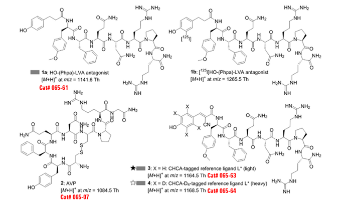
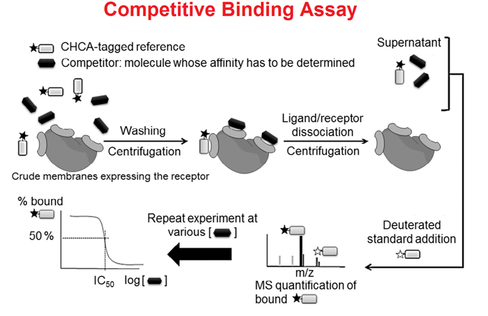
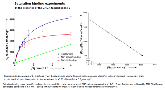
Radiolabeling of ligands is still the gold standard in the study of high-affinity receptor-ligand interactions. In an effort toward safer and simpler alternatives to the use of radioisotopes, we developed a quantitative and highly sensitive matrix-assisted laser desorption ionization mass spectrometry (MALDI-MS) method that relies on the use of chemically tagged ligands designed to be specifically detectable when present as traces in complex biological mixtures such as cellular lysates. This innovative technology allows easy, sensitive detection and accurate quantification of analytes at the sub-nanomolar level. After statistical validation, we were able to perform pharmacological evaluations of G protein-coupled receptor (V1A-R)-ligand interactions. Both saturation and competitive binding assays were successfully processed.
Application of imaging mass spectrometry in drug pharmacokinetics remains challenging due to its weak quantitative capability. Herein, an imaging mass microscope (iMScope), equipped with an optical microscope, an atmospheric pressure ion-source chamber for matrix-assisted laser desorption/ionization (AP-MALDI) and a hybrid quadrupole ion trap time-of-flight (QIT-TOF) analyzer, was first validated and applied to visualize drug disposition in vivo. The distribution and elimination rate of the therapeutic peptide octreotide, a long-acting analogue of the natural hormone somatostatin, was first calculated based on the data determined by iMScope system combining a novel relative exposure strategy. Microspotted pixel-to-pixel quantitative iMScope provided a relative amount of octreotide presented in a thin stomach/intestinal section while preserving its original spatial distribution. The images of dosed mouse stomach clearly demonstrated the transport process of octreotide from the mucosal layer to the muscle side. More importantly, octreotide was found to eliminate from the intestines rapidly, the absorption peak time (Tmax) appeared at 40 min and the half-life time (t1/2) was calculated as 37.7 min according to the elimination curves. Comparisons to the LC-MS/MS data adequately indicated that the quantification approach and methodology based on the iMScope was reliable and practicable for drug pharmacokinetic study.
Matrix-assisted laser desorption/ionization time-of-flight mass spectrometry imaging (MALDI-TOF-MSI) has received considerable attention in recent years since it allows molecular mapping of diverse bimolecular in animal/plant tissue sections, although some barriers still exist in absolute pixel-to-pixel quantification. Octreotide (Cat. #060-25), a synthetic somatostatin analogue, has been widely used to prevent gastrointestine bleeding in the clinic. The aim of the present study is to develop a MALDI-TOF-MSI method for quantitatively visualizing spatial distribution of octreotide in mouse tissues. In this process, a structurally similar internal standard was spotted onto tissue section together with matrix solution to minimize signal variation and give excellent quantitative results. The 2,5-dihydroxybenzoic acid was chosen as the most suitable matrix via comparing the signal/noise generated by MALDI-TOF-MSI after cocrystallization of octreotide with different matrix candidates. The reliability of MALDI-TOF-MSI, with respect to linearity, sensitivity and precision, was tested via measuring octreotide in fresh tissue slices at different concentrations. The validated method was then successfully applied to visualize the distribution of octreotide in mouse tissues after oral administration of octreotide at 20 mg/kg. The results demonstrated that MALDI-TOF-MSI could not only clearly visualize the spatial distribution of octreotide, but also make the calculation of the key pharmacokinetic parameters (Tmax and t1/2) possible. More importantly, the tissue concentration-time curves of octreotide determined by MALDI-TOF-MSI agreed well with those measured based on LC–MS/MS.These findings illustrate the potential of MALDI-TOF-MSI in pharmacokinetic profiling during drug development.
Human proteins can exist as multiple proteoforms with potential diagnostic or prognostic significance. MS top-down approaches are ideally suited for proteoforms identification because there is no prerequisite for a priori knowledge of the specific proteoform. One such top-down approach, termed mass spectrometric immunoassay utilizes antibody-derivatized microcolumns for rapid and contained proteoforms isolation and detection via MALDI-TOF MS. The mass spectrometric immunoassay can also provide quantitative measurement of the proteoforms through inclusion of an internal reference standard into the analytical sample, serving as normalizer for all sample processing and data acquisition steps. Reviewed here are recent developments and results from the application of mass spectrometric immunoassays for discovery of clinical correlations of specific proteoforms for the protein biomarkers RANTES, retinol binding protein, serum amyloid A and apolipoprotein C-III.
Related Products
| Catalog# |
Product |
Standard Size |
Price |
| EK-065-07 |
[Arg8]-Vasopressin (AVP) (Human, Rat, Mouse, Ovine, Canine) - EIA Kit |
96 wells |
$570 |
| 065-07 |
[Arg8]-Vasopressin (AVP) (Human, Rat, Mouse, Ovine, Canine) |
1 mg |
$61 |
| H-065-07 |
[Arg8]-Vasopressin (AVP) (Human, Rat, Mouse, Ovine, Canine) - Antibody |
50 µl |
$225 |
| RK-065-07 |
[Arg8]-Vasopressin (AVP) (Human, Rat, Mouse, Ovine, Canine) - RIA Kit |
125 tubes |
$880 |
| FEK-065-07 |
[Arg8]-Vasopressin (AVP) (Human, Rat, Mouse, Ovine, Canine) - Fluorescent EIA Kit |
96 wells |
$624 |
| EK-065-07CE |
[Arg8]-Vasopressin (AVP) (Human, Rat, Mouse, Ovine, Canine) - EIA Kit, CE Mark Certified |
96 wells |
$595 |
| FEK-065-07CE |
[Arg8]-Vasopressin (AVP) (Human, Rat, Mouse, Ovine, Canine) - Fluorescent EIA Kit, CE Mark Certified |
96 wells |
$649 |
| 065-63 |
CHCA-tagged-LVA antagonist |
100 µg |
$226 |
| 065-61 |
HO-(Phpa)-LVA antagonist |
100 µg |
$226 |
| 065-52 |
pro-Vasopressin (150-164) / Hypothalamic PRP-1 (Human) |
200 µg |
$150 |


Social Network Confirmation